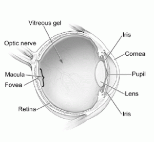
The eye is a very complex organ, with many important parts. To work properly and provide normal vision, every part of the eye must be normal and healthy.
 When you look at something—for instance, a flower—rays of light bounce off the flower and enter the eye through the cornea. The cornea is a clear window on the front of the eye. The light rays pass through the pupil, which is an opening in the middle of the iris. The iris is the part of the eye that determines eye color. Once through the pupil, the light rays are focused by the lens to pass through the clear vitreous gel (which inflates the eye) to reach the retina, where information about the flower is detected and signals are sent through the optic nerve to tell the brain what the flower looks like.
An abnormality of any part of the eye can affect sight. The cornea can become cloudy or irregular, preventing light rays from entering. Dry eye is a common condition that makes the cornea irregular. This causes light rays to bounce off the cornea instead of entering the eye, which reduces vision. Treatments that moisturize the eye, such as lubricating drops or punctual plugs, can smooth out the irregularities and improve vision. Some uncommon problems, such as swelling or infections of the cornea, can make the cornea cloudy, preventing light from entering the eye. If scarring occurs, a corneal transplant is sometimes needed to restore the cornea’s clarity.
The lens of the eye is a clear structure that focuses light on the retina. When we are young, the lens can change its shape to help us focus on objects that are far away or close by. As we reach our 40s, the lens begins to harden and we lose the ability to see up close. Reading glasses or bifocals easily correct this problem. As we get even older, into our 60’s and 70s, the lens becomes cloudy and prevents light from passing through. A cloudy lens is called a cataract. Often cataracts must be removed—and an artificial lens implanted—to restore vision.
Once the light has passed through the cornea and is focused by the lens, it passes through the clear vitreous gel to reach the retina. The retina is the wallpaper that lines the back of the eye, and is like the film in a camera—it is where the picture is made. Once the picture is made on the retina, the optic nerve carries the picture to the brain. All of our fine, straight-ahead vision is handled by the central part of the retina, called the macula. Any condition that affects the macula can reduce vision. Common problems with the macula include macular degeneration, a common condition of aging, and macular swelling, which can occur in diabetics or after cataract surgery. Medications and laser therapy can sometimes improve vision in eyes with disorders of the macula.
The optic nerve carries vision information from the eye to the brain. Problems with the optic nerve can also reduce vision. These include strokes of the optic nerve, as well as inflammation and swelling, which is often seen in people with multiple sclerosis. Because the optic nerve is part of the brain, damage to it is often permanent and results in permanent vision loss.
For more information on Refractive Surgery, click here.